Figure 5.
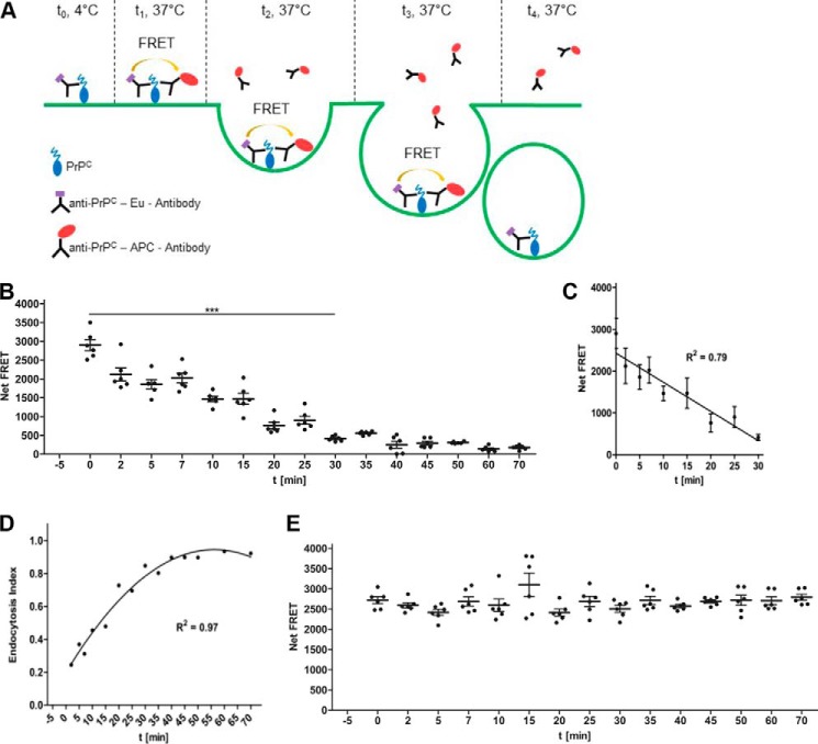
Principle of the endocytosis FRET immunoassay and detection of PrPC uptake by human cells. A, schematic representation of the endocytosis FRET assay. PrPC on the cell surface is first labeled with the donor FRET Eu-labeled antibody (t = 0; 4 °C). After removing excess antibody, cells are exposed to 37 °C to initiate the uptake of Eu-labeled cell-surface PrPC (t = 1). During the course of internalization, Eu-labeled cell-surface PrPC is endocytosed from the cell surface into the cell, and residual Eu-labeled cell-surface PrPC is measured by adding the acceptor FRET antibody at various time intervals (t = 1–4). The endocytosis rate of PrPC is calculated according to the endocytosis index formula (see “Experimental procedures”). B, time course of PrPC endocytosis. A549 cells were first labeled with POM2-Eu at 4 °C followed by the exposure to 37 °C to initiate the internalization of PrPC. At different time points (t = 0–70 min) POM2-APC was added, and the Net-FRET of the remaining cell-surface POM2-Eu-labeled PrPC was measured. C, linear range of PrPC internalization. Error bars represent the standard deviation (±S.D.) of six replicate measurements. D, determination of the internalization rate. The PrPC endocytosis index was calculated according to the formula in “Experimental procedures” and reached a half-life time of ∼25 min. E, time course of cell-surface PrPC A549 cells were labeled with POM2-Eu/POM2-APC at 4 °C followed by exposure to 37 °C. The Net-FRET of cell-surface PrPC was measured at different time points.

