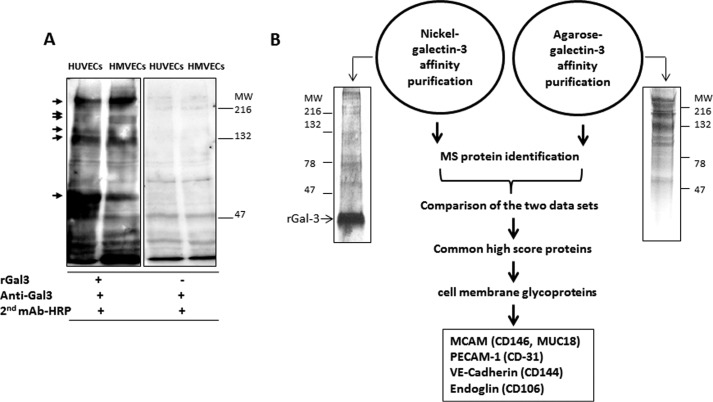
Figure 2.
Identification of endothelial cell surface galectin-3-binding proteins. A, galectin-3 blotting/overlay shows similar binding of galectin-3 to a number of proteins in HUVECs and HUVECs. MW, molecular weight. B, galectin-3 affinity purification and protein identification. HUVEC cell lysates were separated by SDS-PAGE and stained by silver staining (left panel) or applied to nickel- or agarose-conjugated galectin-3 columns. Bound proteins were released by lactose and identified by mass spectrometry or separated by SDS-PAGE and stained by silver staining (right panel).

