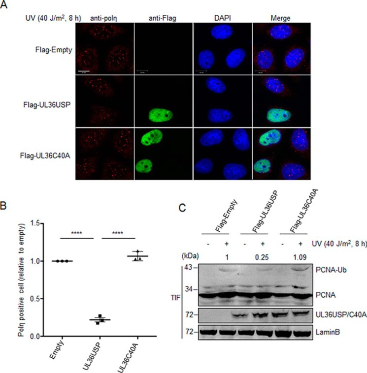
Figure 3.
UL36USP inhibits polη foci formation in response to oxidative DNA damage. A, the number of polη foci was significantly decreases by UL36USP but not C40A. HeLa cells were transfected with UL36USP or -C40A plasmids and subjected to UV treatment. At 30 min after DNA-damage stimuli cells were fixed by cold methyl alcohol, immunofluorescence staining was performed using anti-polη and anti-FLAG antibodies. Endogenous polη were indicated by red fluorescence and UL36USP or UL36C40A by green. The scale bar represents 10 μm. B, quantification of polη foci-positive cells. Percentages of polη foci-positive cells (≥10 polη foci) were counted separately by Bitplane Imaris software. Three independent experiments were performed and ∼1000 cells were counted in each group. Error bars shows S.D. ****, p value < 0.0001. C, PCNA ubiquitination levels decreased in UL36USP-expressing cells compared with the control or UL36C40A group. HeLa cells transfected with UL36USP or -C40A plasmids were treated with or without UV. At 30 min after stimuli, cells were incubated with Triton X-100 buffer. Ubiquitinated PCNA in Triton X-100-soluble and -insoluble fractions was detected separately by Western blotting. The amount of ubiquitinated PCNA was normalized to Lamin B and indicated above the blot.

