Figure 1.

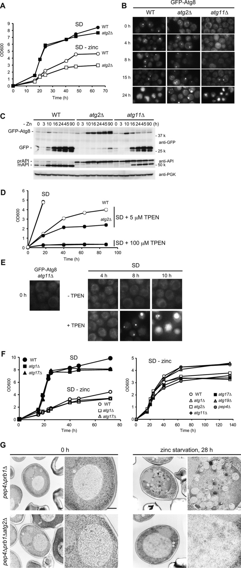
Autophagy is induced upon zinc starvation. A, growth in SD or SD(−zinc). Wild-type or atg2Δ cells were pre-grown in SD to mid-log phase (A600 =1) and then inoculated into fresh SD or SD(−zinc) at A600 = 0.05. Growth was monitored by measuring absorbance at 600 nm. B, GFP-Atg8 localization to the PAS and transport to the vacuole. Wild-type, atg2Δ, or atg11Δ cells expressing GFP-Atg8 were cultured as in A, and GFP-Atg8 was analyzed by fluorescence microscopy. C, processing of GFP-Atg8. Cells were cultured as in B. At the indicated time points, lysates were prepared and analyzed by Western blotting. D, zinc depletion by TPEN. Wild-type or atg2Δ cells were pre-grown in SD to log phase (A600 = 1), and then inoculated into fresh SD with or without TPEN (5 or 100 μm) at an initial A600 of 0.05. At the indicated time points, cell density was measured. E, microscopic observation of GFP-Atg8 expressed in atg11Δ cells in the absence (−TPEN) or presence of TPEN (+100 μm) in SD. F, growth of several types of atg mutants in SD or SD(−zinc). Wild-type, atg1Δ, atg2Δ, atg11Δ, atg17Δ, atg19Δ, or pep4Δ cells were cultured as in A. G, electron microscopic analyses. To observe autophagic bodies in the vacuoles, pep4Δprb1Δ or pep4Δprb1Δatg2Δ cells were used. The cells were cultured in SD(−zinc) for 0 or 28 h and then examined by transmission electron microscopy. Scale bar, 500 nm.
