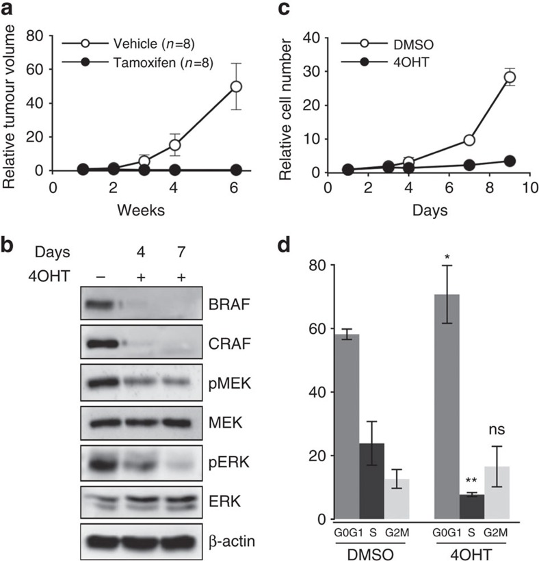
Figure 2. RAF signalling is required for cell proliferation and tumour growth in NRASQ61K-induced murine melanoma.
(a) A melanoma from an untreated Braff/f;Craff/f;Tyr::NRASQ61K/o;Ink4a+/−;Tyr::CreERT2/o mouse was cut into small pieces and subcutaneously grafted into two groups of nude mice that were treated either with tamoxifen or vehicle for 2 weeks. The effect on tumour growth was assessed by measuring tumour volume over a 6-week period. Tumour volumes are plotted relative to the initial volume at the start of treatment. This experiment is representative of three independent experiments requiring 48 Swiss Nu/Nu females (6-week-old) for one primary tumour from a 1-year-old female on a SV129/C57Bl6 mixed genetic background. (b) Western blot analysis of BRAF and CRAF protein levels and MEK and ERK activation levels (pMEK and pERK, respectively) in protein lysates from culture in c on days 4 and 7 of 4OHT treatment compared to DMSO-treated culture. Total MEK, total ERK and β-actin are shown as a loading control. (c) Growth curve analysis of melanoma cell culture established from an untreated Braff/f;Craff/f;Tyr::NRASQ61K/o;Ink4a+/−;Tyr::CreERT2/o primary mouse tumour in response to 4OHT or DMSO for 9 days. Cell number is plotted relative to the initial number of cells at the start of treatment. Data are representative of three independent experiments. (d) Cell cycle analysis by FACS from culture in c on day 6 of 4OHT treatment compared to DMSO-treated culture. Data are the mean value of three independent experiments. *P value <0.05 and **P value <0.01 compared by Student's t-test. ns, not significant. All data are represented as mean±s.d.

