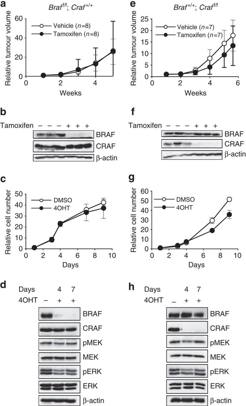
Figure 3. Compensatory functions of BRAF and CRAF for cell proliferation and tumour growth in NRASQ61K-induced murine melanoma.
(a,e) A melanoma from an untreated Braff/f;Craf+/+;Tyr::NRASQ61K/o;Ink4a+/−;Tyr::CreERT2/o or Braf+/+;Craff/f;Tyr::NRASQ61K/o;Ink4a+/−;Tyr::CreERT2/o mouse (a,e, respectively) was cut into small pieces and subcutaneously grafted into two groups of nude mice and experimented as in Fig. 2a. These experiments required 48 Swiss Nu/Nu females (6-week-old) for each primary tumour from a 5-month-old female and a 1-year-old male on a SV129/C57Bl6 mixed genetic background, respectively. (b,f) Western blot analysis for BRAF and CRAF expression at the end of treatment by tamoxifen or vehicle in three individual and representative tumours from a,e, respectively. β-actin is used as a loading control. (c,g) Growth curve analysis of melanoma cell culture established from an untreated Braff/f;Craf+/+;Tyr::NRASQ61K/o;Ink4a+/−;Tyr::CreERT2/o or Braf+/+;Craff/f;Tyr::NRASQ61K/o;Ink4a+/−;Tyr::CreERT2/o primary mouse tumour (c,g, respectively) in response to 4OHT or DMSO for 9 days as in Fig. 2c. (d,h) Western blot analysis of BRAF and CRAF protein levels and MEK and ERK activation levels in protein lysates from culture in c,g respectively, as in Fig. 2b. All data are represented as mean±s.d.

