Figure 5.
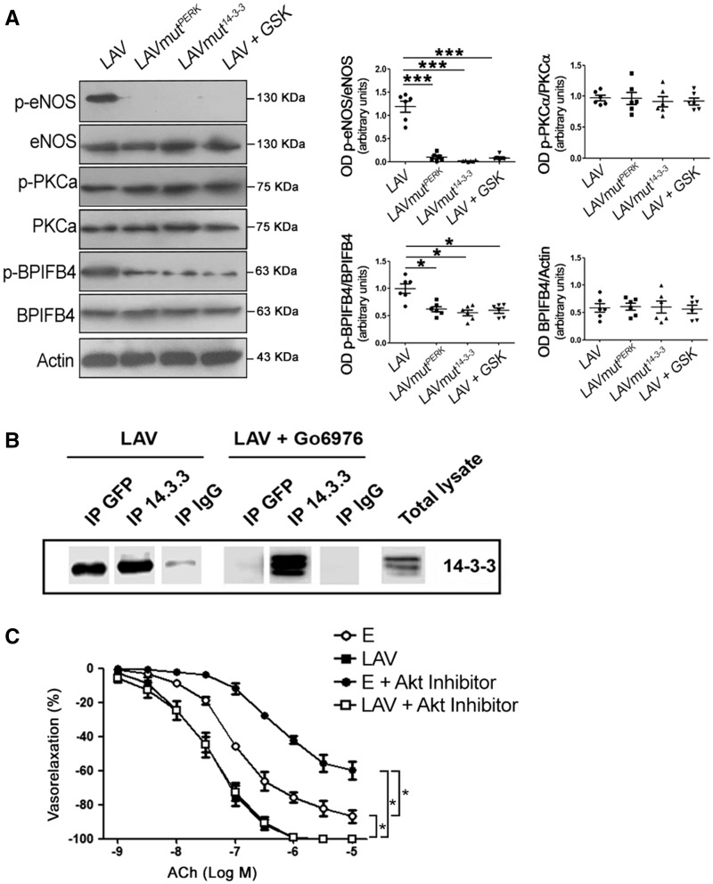
Activation of PKCα is independent of phosphorylation of BPIFB4 by PERK at serine 75. (A) Western blot of ex vivo mouse mesenteric arteries overexpressing LAV-BPIFB4, LAV-BPIFB4mutPERK (Ser75Ala variation), LAV-BPIFB4mut14–3-3 (Ser82Asn variation), or LAV-BPIFB4 plus GSK2606414 (a PERK inhibitor). On the right, graphs of quantification of p-eNOS (S1177), p-PKC-alpha (T497), p-BPIFB4, and BPIFB4. Values are means ± S.E.M., n = 6 experiments. Statistics was performed using one-way ANOVA following Bonferroni’s Multiple Comparison Test; ***P < 0.001 vs. LAV-BPIFB4mutPERK; ***P < 0.001 vs. LAV-BPIFB4mut14–3-3; ***P < 0.001 vs. or LAV-BPIFB4 plus GSK2606414. (B) Co-immunoprecipitation of BPIFB4 and 14-3-3 in HEK293T cells overexpressing LAV-BPIFB4 tagged with GFP protein and treated with the PKCα inhibitor Gö6976. Immunoprecipitation was performed with anti-GFP (directed toward LAV-BPIFB4-GFP), anti-14-3-3, and anti-IgG (as negative control) antibodies followed by immunoblotting with anti-14-3-3 (n = 2 independent experiments). (C) Dose–response curves for ACh of ex vivo C57BL/6 mouse mesenteric arteries transfected with empty vector (E) or with LAV-BPIFB4 in the presence (+) or absence of an Akt inhibitor. Values are means ± S.E.M., n = 6 experiments. Statistics was performed using two-way ANOVA; *P < 0.05.

