
Fig. 6.
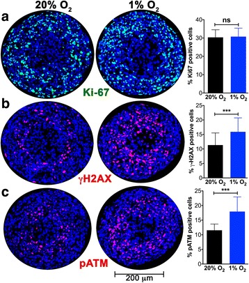
ATM activation and γ-H2AX formation are promoted by hypoxia, while proliferation is unaffected. a The effect of maintaining ~400 μm A673 spheroids in 1% O2 for 12 h was monitored. Spheroid sections were stained for the proliferation marker Ki-67 (green). The % Ki-67-positive cells is plotted (mean and standard deviation from >12 spheroids and two independent experiments). Hypoxia had no effect on cell proliferation. b The DDR marker γ-H2AX (red) is upregulated in the core of spheroids maintained in 1% O2 for 12 h. Bar graph shows the mean and standard deviation from >45 spheroids and >5 independent experiments. c Activation of ATM kinase is indicated by staining for pATM (red) in spheroids maintained in 1% O2 for 12 h. Bar graph shows the mean and standard deviation from >15 spheroids and 4 independent experiments. One way ANOVA with Dunnett’s post-test. ***P < .001, ns P > .05