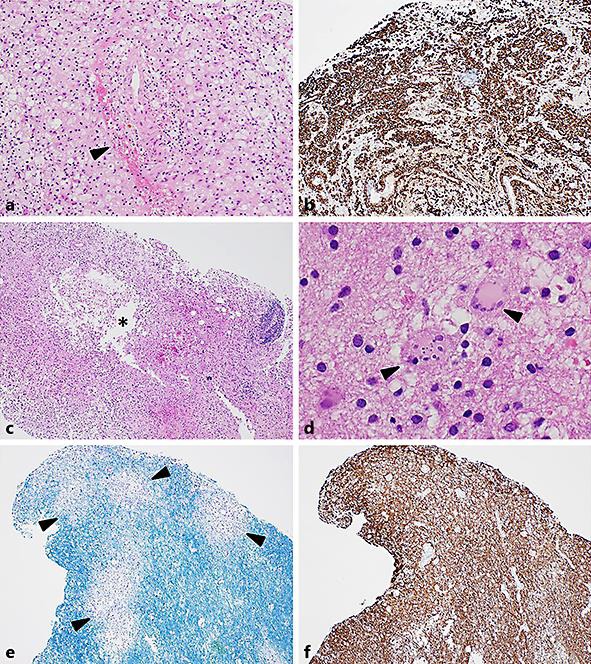
Fig. 2.

Photomicrographs demonstrating the features of the second biopsy. a Perivascular haemorrhage, haemosiderin, and oedema (black arrowhead). There were plentiful foamy macrophages and a mild lymphocytic inflammatory response (haematoxylin and eosin. Original magnification ×100). b Immunohistochemical staining for the macrophage marker CD163, highlighting foamy macrophages as the dominant cell type (CD163 immunoperoxidase, original magnification ×40). c Evidence of early white matter cavitation (asterisk in white space) with a cystic space lined by foamy macrophages. More marked perivascular inflammation present at the biopsy edge (haematoxylin and eosin. Original magnification ×40). d Adjacent gliosis in surrounding white matter containing plump, reactive gemistocytic astrocytes. Scattered astrocytes had fragmented nuclei typical of Creutzfeldt-Peters cells (black arrowheads; haematoxylin and eosin. Original magnification ×400). e, f Serial sections stained with histochemical stain Luxol Fast Blue(LBF)/Cresyl Violet (CV) and immunohistochemical stain for neurofilament (NF), respectively. There was perivascular loss of blue-staining myelin with an irregular margin (black arrowheads). Neurofilament highlighted variable numbers of preserved axons within the areas of myelin loss. At higher magnification occasional axonal spheroids were demonstrated. There was patchy loss of axons within the areas of cystic degeneration (LFB/CV and NF immunoperoxidase, original magnification ×40).
