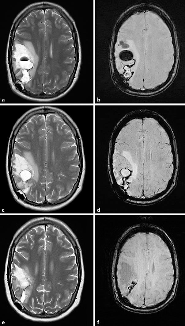
Fig. 3.

Progress magnetic resonance imaging. a T2. b Susceptibility-weighted imaging. Three weeks after presentation, showing a cystic lesion and ongoing surrounding mass effect, consistent with multiple areas of haemorrhagic necrosis. This may represent the natural progression of the disease, though it is difficult to evaluate how much is secondary to postoperative changes. c T2. d Susceptibility-weighted imaging. Five weeks after presentation, showing improving oedema and resolving haemorrhagic cysts. e T2. f Susceptibility-weighted imaging. Two months after presentation, showing significant reduction in the areas of cystic necrosis.
