Fig. 1.

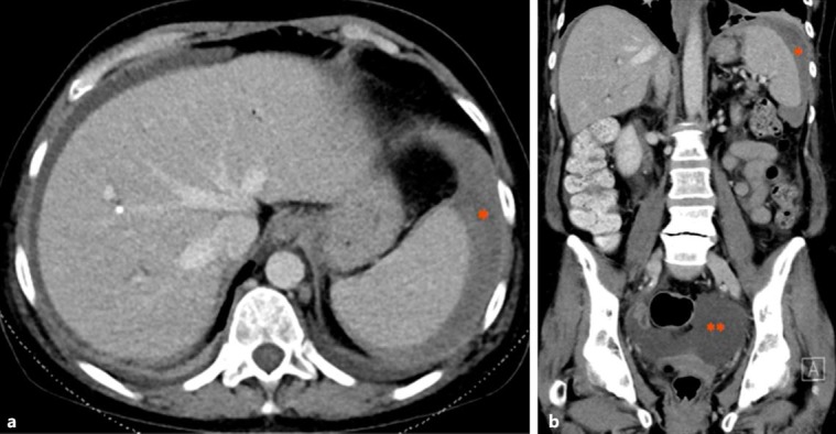
Axial (a) and coronal (b) CT scans in the portal venous phase showing evidence of peri-splenic haematoma (*) and haemoperitoneum (**).

Axial (a) and coronal (b) CT scans in the portal venous phase showing evidence of peri-splenic haematoma (*) and haemoperitoneum (**).