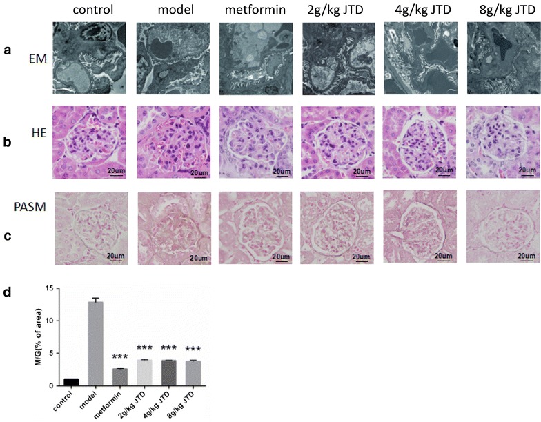
Fig. 3.

JTD alleviates renal histopathology and the ultrastructural pathology of the kidney. a Electron microscopy (EM) analysis of the renal cortex, representative images of glomerular basement membrane thickening and mesangial matrix expansion (scale bar 2 μm; original magnification electron microscopy ×8000; b hematoxylin and eosin (HE) staining of the kidney (original magnification ×400); c periodic schiff-methenamine (PASM) staining of the kidney (original magnification ×400); d JTD lowers the ratio of the mesangial matrix area to the total glomerular area (M/G) via PASM staining. Data are expressed as the mean ± S.D.; N = 5; *P < 0.05; **P < 0.01; and ***P < 0.001 compared to the model group
