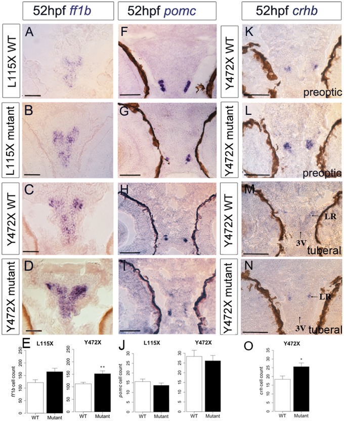
Figure 4.
Abnormal neuronal differentiation in the hypothalamus of disc1 L115X and Y472X embryos. (A–D,F–I) Transverse sections through posterior tuberal hypothalamus at 52 hpf after in situ hybridisation with ff1b (nr5a1a) (A–D) or pomc (F-I). ff1b is expressed more strongly in both L115X (B) and Y472X (D) mutant larvae, compared to wild types (A and C). (E,J) Quantitative analyses of ff1b and pomc cell number at 52hpf. ff1b cell count was not different in L115X embryos (t test, t = −2.45, df = 3.86, P=0.073, N = 3), but was significantly increased in Y472X mutants compared to wild types (t test, t = -3.13, df = 14.06, P=0.007, N = 9–10). pomc cell count was not significantly altered in L115X (t test, t = 1.10, df = 5.56, P=0.318, N = 3–5) or Y472X (t test, t = 0.54, df = 25.38, P=0.593, N = 13–16) embryos (J). (K-O) Transverse sections through preoptic (K,L) or posterior tuberal hypothalamus (M,N) at 52 hpf after in situ hybridisation with crhb in the Y472X line. Quantitative analysis (O) shows significantly more crhb+ cells in the preoptic and tuberal hypothalamus of mutant larvae (t test, t = −2.53, df = 7.83, P=0.036). N = 5 each. Abbreviations: 3V, 3rd ventricle of the hypothalamus; LR, lateral recess of the hypothalamus; WT, wild type larvae; mutant, homozygous mutant larvae. Scale bar: 50 μm.

