Figure 5.
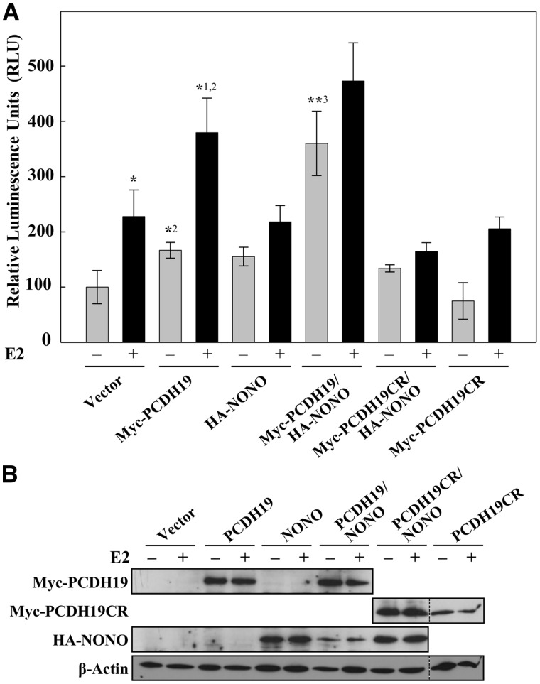
PCDH19 and NONO affect ERα-mediated gene expression. (A) MCF-7 cells were transfected with 3 × ERE TATA luc and control plasmids, wild-type Myc-PCDH19, Myc-PCDH19CR, wild-type HA-NONO, Myc-PCDH19/HA-NONO or Myc-PCDH19CR/HA-NONO expression vectors. Cells were cultured in charcoal-stripped FCS for 16 h and then for 6 h in the presence or absence of 10 nM E2. Data are expressed as relative luciferase activity ± SD from three or more independent experiments. *P < 0.05 comparing +E2 versus –E2 vector control, *1P < 0.05 comparing +E2 versus –E2 WT, *2P < 0.01 comparing +E2 WT versus +E2 vector and –E2 WT versus –E2 vector, **3P < 0.01 comparing –E2 WT/NONO versus –E2 WT using Bonferroni adjusted planned comparisons. (B) Western blot of MCF-7 cell extracts expressing wild-type Myc-PCDH19 or Myc-PCDH19CR were probed with anti-Myc antibody. Transfected HA-NONO was detected using anti-HA antibody while endogenous NONO using anti-NONO antibody. β-Actin was used as a loading control. Figure 5B was generated by cropping full-length western blots shown in Supplementary Material Figure S10. Dotted lines separate the cropped images from different western blots. As HA-NONO and Myc-PCDH19CR were detected at approximately the same size, different immunoblots were used to visualize the two proteins.

