
Fig. 3.
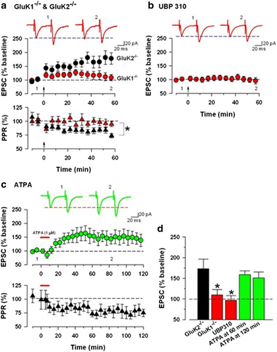
KA receptors mediate the induction of pre-LTP. a Upper panel: In GluK1-/- mice, sample traces of eEPSCs with paired-pulse stimulation at 50 ms during baseline (1) and 60 min after the induction stimulus (2) at a holding membrane potential of -60 mV. Middle panel: GluK2-/-mice showed normal pre-LTP (black circle). GluK1-/- mice showed greatly reduced pre-LTP (red circle. Bottom panel: PPR for the GluK2-/- (black) and GluK1-/-(red) groups. b A specific GluK1 antagonist, UBP310 (10 μM), completely blocked pre-LTP. c Upper panel: A GluK1 agonist, ATPA (1 μM for 10 min), induced long lasting potentiation, recorded for 2 h. Bottom panel: Averaged data of PPR change before and after ATPA application. d Summary of the effects of GluK2-/-, GluK1-/-, a GluK1 antagonist or a GluK1 agonist on pre-LTP. The amplitudes of eEPSCs in GluK1-/- or UBP310 groups were significantly decreased compared with control pre-LTP. There was no difference among control pre-LTP, GluK2-/- and ATPA. Modified from Koga et al [38]