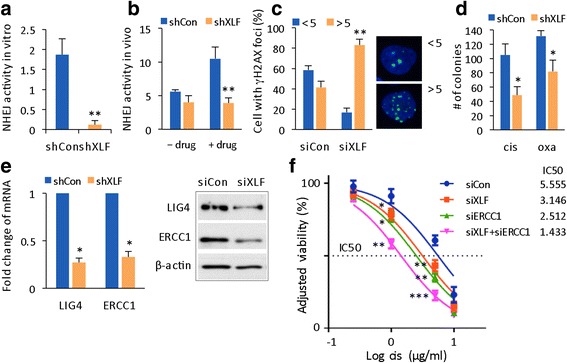
Fig. 3.

Knockdown of XLF chemosensitizes resistant 97 L cells by causing inhibition of NHEJ activity. a In vitro NHEJ activity was assayed and quantified in shRNA-Con and shRNA-XLF lentivirus-infected 97 L cells from 3 independent experiments. b In vivo NHEJ activity was calculated as the percentage of GFP-positive cells and normalized to transfection efficiency for the shCon and shXLF group, which were transfected with pEGFP-PEM1-Ad2 plasmid and treated with or without oxaliplatin. The data are reported as the mean ± SD, n = 4. c Quantification of the percentage of cells with <5 and >5 γΗ2ΑX foci per nucleus at 26 h after drug treatment for the siRNA-Con and siRNA-XLF groups (left panel). The data are reported as the mean ± SD, n = 2. Image of γH2AX speckles with <5 and >5 foci per nucleus (right panel). d Statistical comparison of drug sensitivity based on colony formation assay results from shCon and shXLF cells. Number of colonies is reported as the mean ± SD from 2 independent experiments. e Gene expression levels (left panel) and protein levels (right panel) of LIG4 and ERCC1 were determined by qRT-PCR and WB, respectively, in shCon and shXLF HCC cells. f Cisplatin response curves and IC50 concentrations for siCon, siXLF, siERCC1, and siXLF + siERCC1 cells, respectively. Statistical comparison was between siXLF, siERCC1, and siXLF + siERCC1 versus siCon, respectively, with treatment by cisplatin 1 or 5 μg/ml. * p < 0.05; ** p < 0.01; *** p < 0.001
