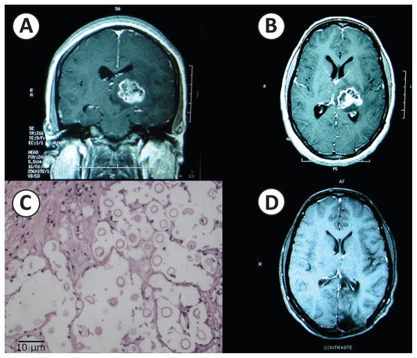
FIGURE 1.
Central nervous system cryptococcoma. T1-weighted post-gadolinium coronal (A) and transverse (B) MRI images revealing a 3cm x 2cm enhancing mass in the left thalamus with central hypointensity suggestive of necrosis. C: Hematoxylin and Eosin stain of brain biopsy tissue at 40x revealing numerous budding encapsulated organisms consistent with Cryptococcus species. D: T1-weighted post-gadolinium transverse magnetic resonance imaging (MRI) image revealing post-craniotomy changes but no evidence of an intra-parenchymal mass.

