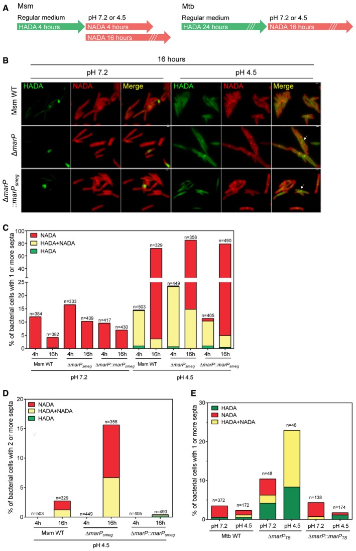
Figure 2. The separation of progeny cells is impaired in ΔmarP mutants during acid stress.

- Schematic of peptidoglycan labeling experiments for Msm and Mtb.
- Representative images of Msm WT, ΔmarP, and ΔmarP::marP smeg incubated in 7H9 medium containing 1 mM HADA for 4 h, washed, and further incubated in Sauton's medium at pH 7.2 or at pH 4.5 containing 1 mM NADA for 16 h. Arrows show examples of septa labeled with both HADA and NADA. Scale bars, 1 μm. See also Fig EV1B.
- Proportion of bacteria that contained at least one septum. All septa labeled with HADA only (green), with NADA only (red), or with at least one septum labeled with both probes (yellow) are reported for Msm WT, ΔmarP, and ΔmarP::marP smeg. The bacteria were incubated in 7H9 medium containing 1 mM HADA for 4 h, washed, and further incubated in Sauton's medium at pH 7.2 or pH 4.5 containing 1 mM NADA for 4 or 16 h.
- Proportion of bacteria in (C) that contained at least two septa after incubation in Sauton's medium at pH 4.5 for 4 or 16 h. Green, red, and yellow indicate bacteria for which all septa were labeled with HADA only, NADA only, or that had at least one septum labeled with both probes, respectively.
- Proportion of bacteria with a septum labeled with HADA only (green), NADA only (red), or both HADA and NADA (yellow). Mtb WT, ΔmarP, and ΔmarP::marP TB cells were incubated in 7H9 medium containing 1 mM HADA for 24 h, washed, and further incubated in Sauton's medium at pH 7.2 or at pH 4.5 containing 1 mM NADA for 16 h. See also Fig EV1C.
