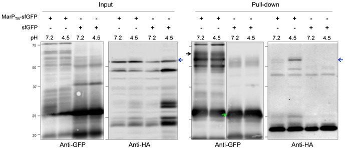
Figure EV3. RipATB and MarPTB interact specifically at acidic pH (related to Fig 5).

Co‐immunoprecipitation from whole‐cell protein lysates of Msm ΔmarP::MarP TB ‐sfGFP::RipA TB ‐HA and ΔmarP::sfGFP::RipA TB ‐HA incubated for 4 h at pH 4.5 or pH 7.2. MarP‐sfGFP and sfGFP were pulled down using anti‐GFP‐coated magnetic beads. Whole‐cell lysates (input) and GFP eluates (pull‐down) were analyzed by SDS–PAGE, and MarPTB‐sfGFP (black arrow), sfGFP (green arrow), and RipATB‐HA (full‐length RipA, blue arrows) were detected by Western blot using anti‐GFP or anti‐HA antibodies. Data are representative of two independent experiments.Source data are available online for this figure.
