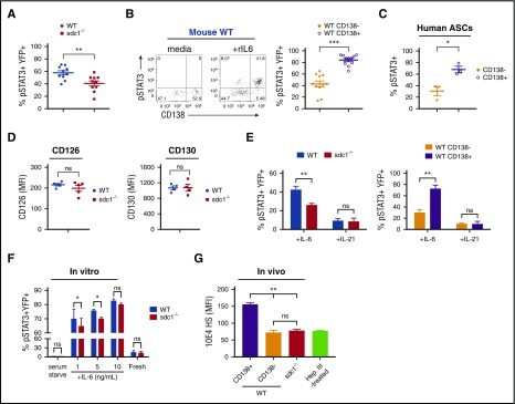
Figure 4.
CD138 expression on ASCs promotes IL-6 signaling. (A-B,D) Mice were co-injected with purified transgenic B cells from WT and sdc1−/− mice, immunized, and examined for YFP+ ASCs on day 7. (A) dLN cells were briefly serum starved, incubated in the presence of recombinant IL-6 (rIL-6) for 20 minutes and analyzed for intracellular pSTAT3 by flow cytometry. Graph represents mean (± SEM) of YFP+pSTAT3+ cells. (B) Within the WT ASC compartment, mature CD138+YFP+ ASCs were distinguished from immature CD138−YFP+ ASCs by flow cytometry, and stained for pSTAT3 following ex vivo stimulation by rIL-6. Representative flow cytometry dot plots depict prior to (media) and after (+rIL6) rIL-6 stimulation (left). Graph represents mean (± SEM) of the percentage of YFP+pSTAT3+ cells from each subset (right). (C) Human PBMCs were examined by flow cytometry. Graph represents mean (± SEM) of the percentage of CD19lowCD38high ASCs positive for pSTAT3 from within the CD138+ or CD138− subsets following ex vivo stimulation by human rIL-6. (D) The dLN from immunized host mice were examined by flow cytometry. YFP+ ASCs derived from either WT or sdc1-deficient B cells were examined for IL-6α receptor (CD126; left) or IL-6β (CD130; right) receptor expression. Graphs show mean of MFI from either WT (blue) or sdc1−/− (red) cells. (E) dLN cells from immunized mice were briefly serum starved, incubated in the presence of rIL-6 or rIL-21 (10 ng/mL) for 20 minutes, and analyzed for intracellular pSTAT3 by flow cytometry. Graphs show mean (± SEM) of the percentage of YFP+ cells stained for pSTAT3. (F) Purified B cells from WT and CD138-deficient mice (sdc1−/−) were cocultured with LPS to generate ASCs in vitro. On day 3, cells were incubated in media in the presence (fresh) or absence (serum starved) of fetal calf serum, and stimulated with rIL-6 (either 1, 5, or 10 ng/mL). pSTAT3 levels were examined by flow cytometry. Graph depicts mean (± SEM) of the percentage of YFP+pSTAT3+ cells. (G) In vivo generated YFP+ ASCs were stained with 10E4 antibody to detect HS. Heparinase III-treated cells that lack HS surface expression is used as a control. Graph represents the MFI of 10E4 expression on various subsets relative to heparinase III-treated cells. *P < .05; **P < .01; ***P < .001. ns, not significant; PBMCs, peripheral blood mononuclear cells.

