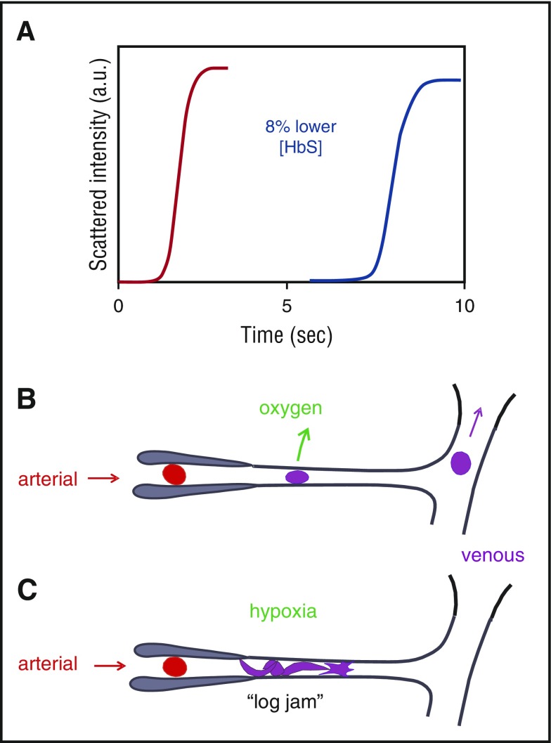
Figure 1.
Connection between kinetics and pathophysiology. (A) Schematic of kinetic progress curve for polymerization occurring on the seconds time scale measured by light scattering due to fiber formation. Before the appearance of fibers, there is a delay (lag phase).38 The delay time is extraordinarily sensitive to HbS concentration, depending on the 30th power of the concentration.38,40 Such a large exponent means that a decrease of only 8% in the HbS concentration increases the delay time 10-fold. (B) Schematic of microcirculation: arteriole, capillary, and venule. The vast majority of cells escape the microcirculation before fibers form and cause cellular distortion (sickling).34,41 (C) Schematic of vaso-occlusion. If the delay time is shorter than the transit time (or fibers have not completely dissolved upon oxygenation in the lungs42 and can grow without a delay34,43), fibers form within the small vessels and can cause vaso-occlusion. In this graphic, factors that slow the transit of red cells through the microcirculation, such as increased adherence to the vascular endothelium by damaged red cells or increased leukocytes associated with infection will increase the probability of vaso-occlusion. a.u., arbitrary units.

