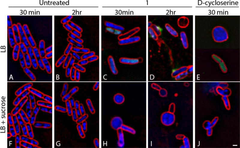
Figure 5.

Effects of compound 1 on cell wall biosynthesis in E. coli ΔtolC. (A, B, F, G) Untreated cells. (C, D, H, I) Cells treated with compound 1 for either 30 minutes or two hours at 5 × MIC (25 μg/mL). (E, J) Cells treated with ᴅ-cycloserine at 5 × MIC (125 μg/mL) for 30 minutes. Cells (F–J) were treated in the presence of 0.5 M sucrose to facilitate visualization of cell shape defects. Cells were stained with FM 4–64 (red), DAPI (blue), and SYTOX Green (green). Scale bar is 1 μm.
