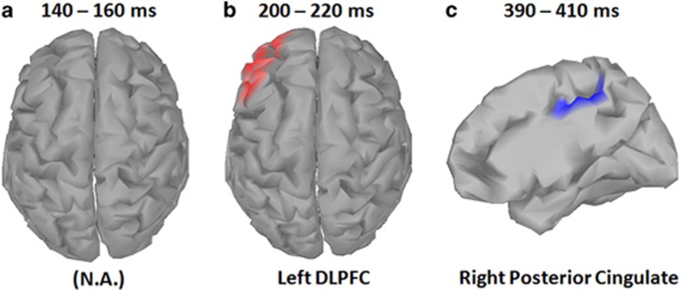
Figure 5.
Electroencephalographic (EEG) source analysis for target trials. Cortical maps indicate sources for event-related potential (ERP) peak activation during each time window. No difference between schizophrenia patients and healthy comparison participants were observed early time window (P1; a). Source localization analyses of the middle time window (N2) indicated significantly greater positive source activity (current source/efferent dipole), in the dorsolateral prefrontal cortex (DLPFC) for healthy participants compared to schizophrenia patients (b). Source localization analyses also indicated greater negative source activity (current sink/afferent dipole) in the right posterior cingulate cortex in the later time window (P3) for healthy participants compared with schizophrenia patients (c). Source analyses were computed for the 20 ms (10 ms pre and post) surrounding the group-level ERP peak amplitude for that time window. For both N2 and P3 windows, healthy participants demonstrated greater source activity compared to schizophrenia patients, with source colors representing dipole direction of significant between group differences. A full color version of this figure is available at the Neuropsychopharmacology journal online.

