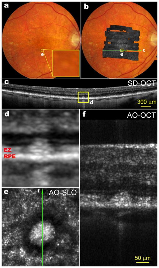
Figure 7.
Multimodal imaging of SDD. (a), Color fundus photograph. The yellow box (e) is of 300 μm on a side, (b), AOSLO montage overlaid on the fundus photograph. (c), A SD-OCT B-Scan taken along the green arrow-line in panel b shows that this SDD has broken the photoreceptor EZ band. (d), Magnification of boxed area in panel c. (e), The AOSLO image of the boxed retina in panels a and b. The bright spots outside the hyporeflective annuls are photoreceptors, mostly cones. (f), The AO-OCT scan of the SDD, as indicted by the green arrow line in panel e. The scale bar in panel f also applies to panels d and e. All SD-OCT images are in logarithmic grey scale. AO-OCT is in linear grey scale. The subject is an 83-year-old non-Hispanic man of European-descendant with intermediate stage non-neovascular AMD. (From Y. Zhang, unpublished)

