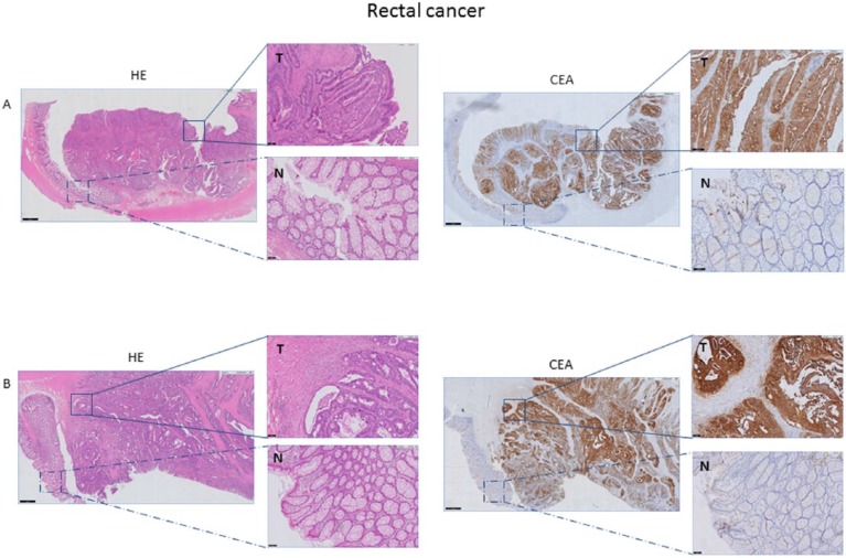
Figure 2.
Representative example of HE and CEA staining of rectal cancer tissue. CEA expression of tumor tissue (T) compared with adjacent normal (N) rectal tissue, derived from a patient with a preoperative serum CEA level <3gn/mL (A) and >3 ng/mL (B) magnification 1x and 10x). Both tumor tissues (A and B) show an intense, circumferential CEA expression, independent of the preoperative serum CEA level. Normal epithelium shows weak expression. CEA indicates carcinoembryonic antigen; HE, hematoxylin-eosin.

