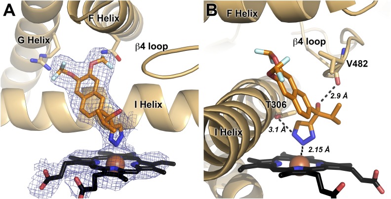
Fig. 6.
The 3.1 Å structure of CYP17A1 with (S)-seviteronel. (A) (S)-seviteronel (orange sticks) binds in the CYP17A1 active site coordinated to the heme (black sticks) iron (red sphere). Electron density for ligand and heme (blue mesh) is a simulated annealing 2Fo − Fc composite omit map contoured to 1.00 σ. B) Interactions between CYP17A1 active site and (S)-seviteronel. Distances shown are averages from all four molecules.

