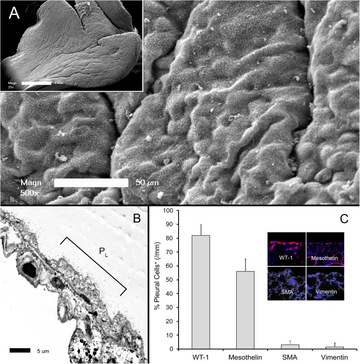
Fig 1. Morphology of the visceral pleural mesothelium.
A) Scanning electron microscopy (SEM) of the cardiac lobe 3 days after sham thoracotomy. The pleural surface demonstrated the characteristic "bumpy" appearance of the mesothelium covered by microvilli (inset, whole lobe). B) The thin mesothelial monolayer and microvilli were apparent on transmission electron microscopy (TEM)(PL = free pleural surface). C) The control mesothelium expressed WT-1 and mesothelin; only rare cells expressed the cytoskeletal proteins SMA and vimentin (inset, immunofluorescence staining). Immunostaining reflects the percentage of positive pleural cells per mm of cardiac lobe pleura.

