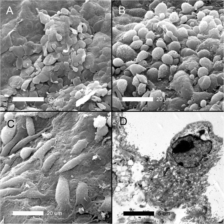
Fig 4. Electron microscopy of the cardiac lobe pleural mesothelium 3 days after pneumonectomy.
A) Cells apparently in the early stages of transition—with disruption of intercellular junctions and loss of microvilli—retained the typical “flagstone” mesothelial morphology. B) Cells apparently in the later stages of transition demonstrating few intercellular junctions and rounded morphology. C) Spindle cell or fusiform morphotypes were found in some regions associated with exposed basement membrane. D) Transmission electron microscopy demonstrated mesothelial cells with Golgi and mitochondrial ultrastructure suggesting significantly enhanced metabolic activity. In some cells, TEM demonstrated chromatin condensation suggesting apoptosis in a subset of pleural cells (not shown).

