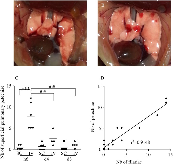
Fig 2. Hemorrhages in the lung of infected mice.
BALB/c mice were inoculated with 40 L3 of L. sigmodontis either subcutaneously (SC) or intravenously (IV). Representative picture of (A) a normal lung, (B) a lung with superficial numerous roundish well-delineated red hemorrhages. (C) Number of superficial pulmonary hemorrhages in lungs at six hours (h6), four days (d4) and eight days (d8) post inoculation. n = 6, bars represent the mean ± SEM; two-way ANOVA followed by Bonferonni, *** = p<0.001 (difference between IV- and SC-infected mice), ## = p<0.01(difference between timepoints in IV-infected mice). (D) Correlation test (Pearson) between the number of L3 recovered in the lung and the number of hemorrhages, r2 = 0.9148.

