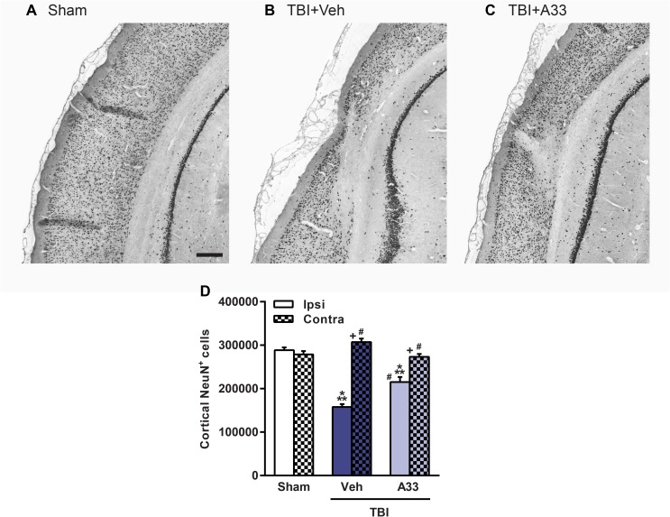
Fig 11. A33 treatment reduced neuronal loss in the pericontusional cortex at 2 months post-injury.
Representative images of the ipsilateral parietal cortex of (A) sham, (B) vehicle-treated and (C) A33-treated TBI animals, immunolabeled for mature neurons using NeuN. Representative images at -5.3 mm bregma, scale bar 250 μm. (D) Quantification of NeuN+ cells in the ipsilateral and contralateral parietal cortex. The number of NeuN+ cells were significantly reduced in the ipsilateral parietal cortex in both vehicle and A33-treated TBI animals as compared to sham animals. A33 treatment rescued neuronal loss in the pericontusional cortex as compared to vehicle-treated TBI animals. (main effect of treatment: F(2, 64) = 19.21, p<0.0001; main effect of region (ipsi vs. contra): F(1, 64) = 93.68, p<0.0001; interaction of treatment x region: F(2, 64) = 43.89, p<0.0001.) Mean ± SEM, n = 10-13/group, ***p<0.001 vs. Ipsi/Contra Sham, #p<0.001 vs. Ipsi TBI+Vehicle, +p<0.001 vs. Ipsi TBI+A33, two-way ANOVA with post-hoc Student-Newman-Keuls.

