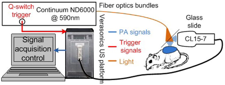
Fig. 2.

Experiment setup for xenograft tumor imaging in vivo. 590 nm laser targeting the optical absorption peak of Coomassie Blue encapsulated in the NPs were used. The laser was delivered through a fiber optics bundle. A US probe acquires US and PA measurements, which is processed and displayed by the Verasonics US platform [10].
