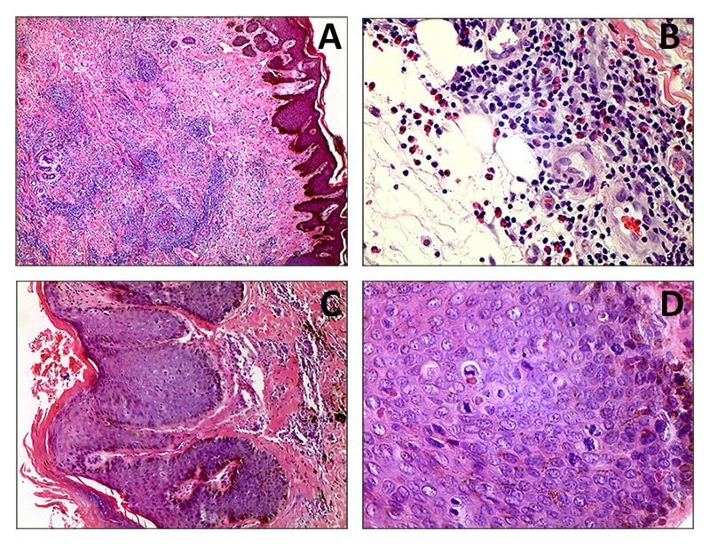
Figure 3. A (4x) y B (40x). A malignant neoplasm lesion of lymphoid origin formed by groups of cells with monotonous, small-lobed nuclei infiltrating into the dermis, accompanied by a moderate number of eosinophils. In some areas, hyperplastic epithelium with Pautrier microabscesses can be observed. C (10X) y D (40X). Skin compromised by a malignant neoplasm lesion of epithelial origin, formed by papillomatous epidermal acanthosis, with orthokeratotic hyperkeratosis and infiltrating tracts of epidermoid differentiated cells. These infiltrated cells possess ovoid nuclei, some with dispersed chromatin and some presenting anisonucleosis, perinuclear cytoplasmic vacuolization and possible Bowenoid transformation, which may suggest a viral etiology. The mitotic activity of the lesion is frequent. Adjacent stroma presented with moderate chronic inflammatory infiltrate.