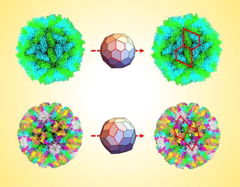
Key Figure, Figure 3. Chiral Pentagonal Frameworks Describe the L-A Virus and Polyomavirus Capsids.
The top images show how the pentagonal hexecontahedron is able to accurately capture the geometry of the L-A virus capsid (PDB ID: 1M1C) by superimposing two appropriate pentagons (outlined in red) around a two-fold axis of the assembly. Similarly, the bottom images show two units of the asymmetric pentagonal hexecontahedron superimposed on the polyomavirus capsid (PDB ID: 1SIE). In both cases, the pentagons capture the chirality and outline key viral protein subunit interfaces, thereby providing a more fitting description than triangles. The calculated dimensions of the appropriate pentagons are given in Supp. Figures 5 and 8.

