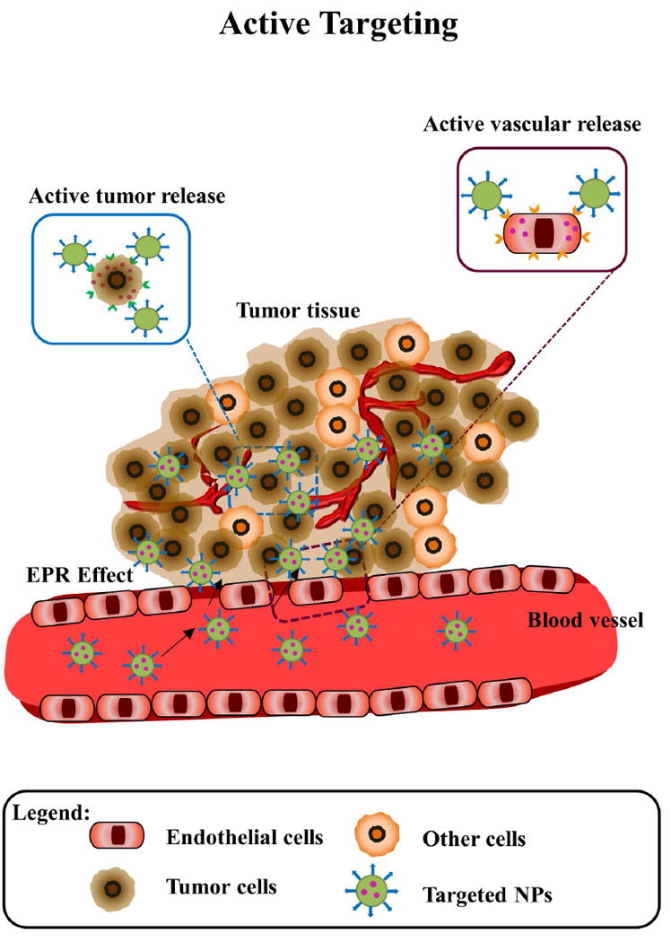
Figure 4. Schematic of active (targeted) nanoparticles in cancer.

Nanoparticles extravasate from the vasculature due to EPR effect, then targeted nanoparticles bind and internalize into tumor tissues, the retention and uptake of these nanoparticles in cancer cells (brown) is augmented due to specific antigen-antibody/ ligand-receptor interactions, and wash out of nanoparticles is reduced.
