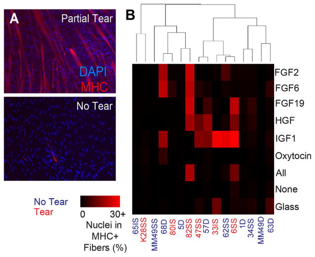
Figure 3.
SMP differentiation capacity varies by tear state and proliferation medium. (A) Cells were seeded at high confluency at the end of the proliferation experiment (P7-8) and allowed to differentiate for 5 days in differentiation medium (5% horse serum and 10 μg/ml insulin). Representative images of cells are shown from partial tear and no tear. (B) Differentiation was quantified as the number of nuclei that were in myosin heavy chain (MHC)-positive myotubes. Growth factor effect p = 0.00698, tear effect p < 10−4, growth factor*tear interaction p = 0.00547. Data were analyzed using a two way ANOVA with Tukey post-hoc analysis. (C) RNA expression of myogenic markers MyoD and Pax7 shown as a ratio of MyoD/Pax7 expression after cell expansion. Error bars are SEM.

