Figure 4.
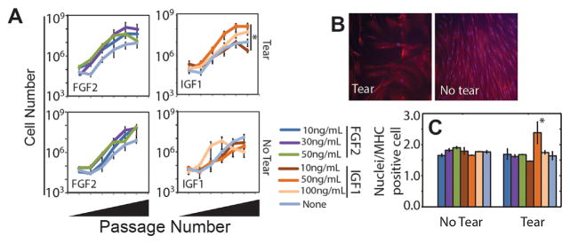
Growth factor dose effects. (A) SMPs were expanded on 11 kPa polyacrylamide gels, laminin-111, collagens type I and IV in the presence of the indicated growth factors and their concentrations. Data are plotted as total cell number versus passage number. n = 3 technical replicates with one biological replicate per tear state. (B) Immunofluorescent staining for MHC from SMPs with the indicated tear state. (C) Number of nuclei per MHC positive cell plotted for the growth factors and concentrations indicated in panel A. *p < 0.05 for comparisons to all other conditions using a post-hoc Tukey test.

