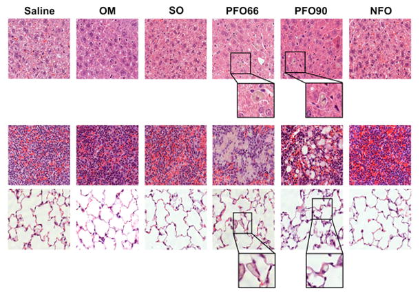
Figure 3. PFO66 and PFO90 result in histologic abnormalities in liver, spleen, and lungs, while NFO preserves normal organ architecture.

Saline, OM, SO, and NFO groups demonstrated histologically normal livers (top row), spleens (middle row), and lungs (bottom row) after 19 days of treatment. PFO66 and PFO90 groups developed hepatic and splenic lipid-laden macrophages without evidence of inflammation or necrosis; as well as pulmonary fat deposits. All panels are stained with hematoxylin and eosin. Magnification is 400X for all groups. For PFO66 and PFO90 groups, a portion of hepatic and pulmonary slides further magnified to emphasize lipid-laden macrophages and pulmonary fat.
