
Fig. 5.
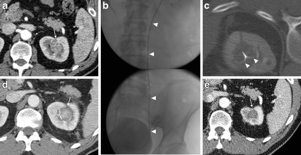
Treatment of a left kidney central tumour with renal sinus extension. a contrast enhanced CT demonstrating a centrally located tumour (white arrow) with extension in close proximity of the renal sinus. b retrograde pyeloperfusion was performed through a single j stent (arrowheads) placed endoscopically the day of the ablation. c CT scan after insertion on a radiofrequency electrode (arrowheads) into the tumour. d contrast enhanced CT performed immediately after the ablation demonstrated the complete ablation of the tumour (white arrowhead) without complications. e Contrast enhanced CT 2 years after the ablation demonstrating complete ablation with tumour shrinkage and no complications