
Figure 3.
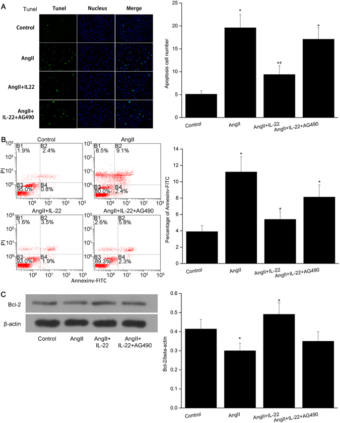
IL22 inhibited the apoptosis of PMVECs induced by AngII. (A) IL22 could attenuate the apoptosis of PMVECs significantly as revealed by TUNEL assay, while such phenomenon was completely inhibited after AG490. (B) Flow cytometry indicated the apoptosis rate of the PMVECs in the AngII+IL22 group was significantly decreased compared with the AngII group. The apoptosis rate in the AngII+IL22+AG490 group was significantly elevated compared with that of AngII+IL22 group. The raw data were listed in the Supplementary file. (C) The expression of Bcl-2 in the PMVECs significantly decreased after AngII stimulation, while the expression of Bcl-2 in the AngII+IL22 group was higher than that of AngII group. *P < 0.05 versus control group. #P < 0.05 versus AngII group and AngII+IL-22+AG490 group. Immunofluorescence images were observed under a magnification of 200×.