
Figure 4.
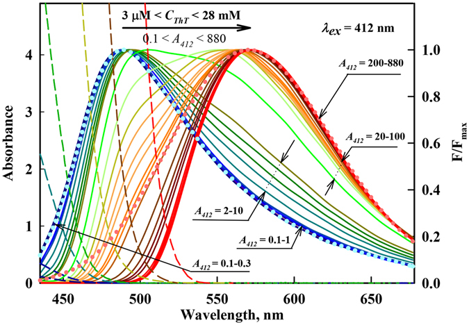
The fluorescence spectra of aqueous solutions of ThT at different concentrations with corrections for the primary inner filter effect and normalization at the spectral maxima. The ThT concentration was increased from 3 μM to 28 mM, which corresponds to an absorbance change from 0.1 to 880. λ ex = 412 nm. The forms of the monomer and excimer fluorescence spectra are given in blue and pink dots, respectively. The dashed curves represent the long wavelength edge of the absorption spectra of ThT solutions with concentrations 0.006, 0.016, 0.11, 0.32, 1.1, 6, 28 mM. Curves with А 412 = 0.1–0.3 correspond to ThT concentrations 3, 6, 10 µM, curves with А 412 = 0.1–1 correspond to ThT concentrations 3, 6, 10, 16, 22, 29 µM, curves with А 412 = 2–10 correspond to ThT concentrations 0.06, 0.11, 0.19, 0.22, 0.32 mM, curves with А 412 = 20–100 correspond to ThT concentrations 0.6, 0.8, 1.1, 1.4, 1.7, 1.9, 2.2, 2.5, 2.8 mM, curves with А 412 = 220–880 correspond to ThT concentrations 6, 13, 19, 25, 1.7, 28 mM.