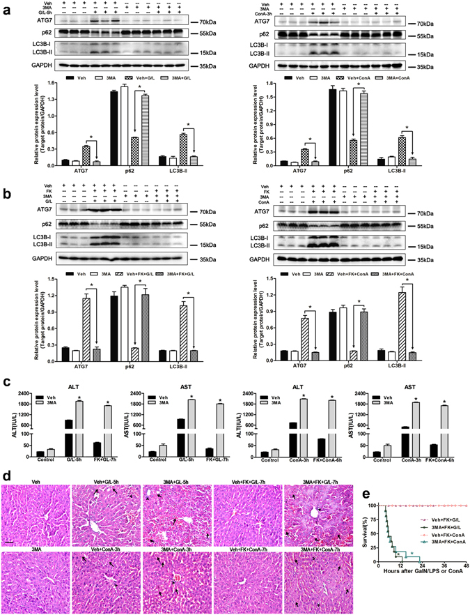
Figure 2.

Autophagy inhibition by 3MA abolishes the FK866-conferred hepatoprotective effect in mice. Mice were injected with 3MA (30 mg/kg, 30 min, IP) prior to GaIN/LPS (G/L, 600 mg/kg /0.5 μg/kg, IP) or ConA (20 mg/kg, IV) challenge in the presence or absence of FK866 (FK, 10 mg/kg, IP). (a) Measurement of autophagy indicators in mice liver. The data are shown as the means ± SEM. n = 4–6 per group. *P < 0.05 indicates significant differences. (b) Western blot analysis of ATG7, p62, and LC3B protein expression in the presence or absence of 3MA. The data are shown as the means ± SEM. n = 4–6 per group. *P < 0.05. (c) Determination of serum ALT and AST levels. The data are shown as the means ± SEM. n = 6 per group. *P < 0.05 vs. the corresponding vehicle (Veh) group. (d) Representative images of liver injury. The arrows denote hepatocellular necrosis; the arrowheads denote infiltrating inflammatory cells. Original magnification × 400, scale bar 50 µm. n = 4 per group. (e) Survival rate of mice after G/L or ConA with FK in the presence or absence of 3MA. n = 11 per group. *P < 0.05 compared to the corresponding Veh-treated group.
