Figure 2.

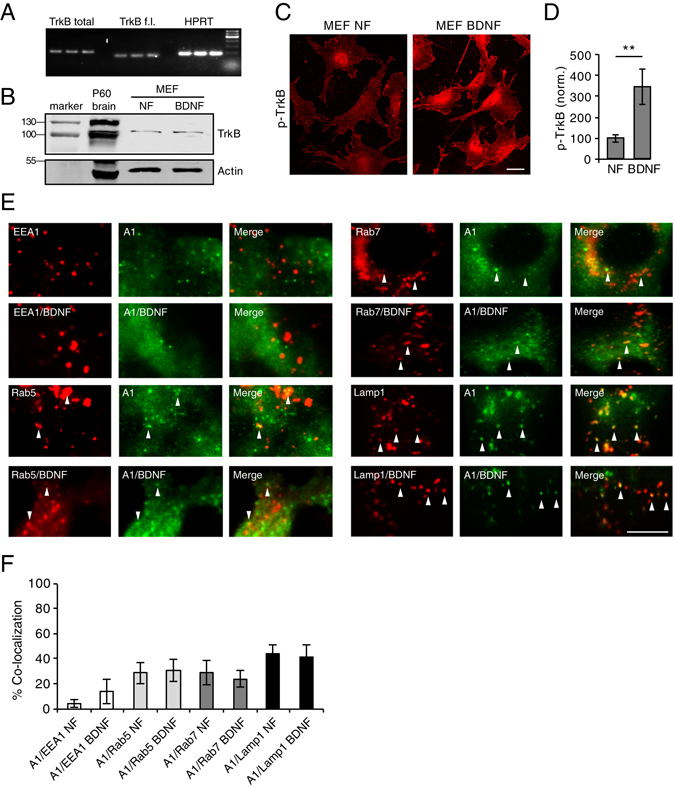
EndophilinA1 colocalizes with endosomes. (A) qPCR products of total TrkB (including full length and truncated isoforms), full length TrkB (TrkB f.l.) and HPRT as a control from mouse embronic fibroblast (MEF) lysates. (B) Western blot of TrkB in MEF lysates in control no factor (NF) conditions, and following treatment with BDNF. P60 brain lysate serves as a positive control, and actin as a loading control. (C) Images of pTrkB immunostaining in cultured MEFs in “no factor” (NF) untreated conditions, or following treatment with 100 ng/ml BDNF for 30 min; scale bar = 10 µm. (D) Quantitation of pTrkB signal in MEFs in NF and BDNF conditions; n-21 images per condition, error = SEM, significance determined by Student’s t-test. (E) Representative TIRF microscopy images of MEFs co-transfected with GFP-tagged EndophilinA1 and the RFP-tagged endosomal markers EEA1, Rab5, Rab7 or Lamp1, in the presence and absence of 100 ng/ml BDNF. Arrows indicate colocalization of EndophilinA1 (green) with endosomal markers (red). (F) Quantitation of colocalization of transfected GFP-tagged EndophilinA1 with RFP-tagged EEA1, Rab5, Rab7, or Lamp1 in control conditions with no factors (NF) or following addition of BDNF; n = 8–10 images per condition from 3 independent cell cultures, significance determined by unpaired two-tailed Student’s t-test comparing non-stimulated to stimulated conditions, error = SEM, scale bar = 10 µm.
