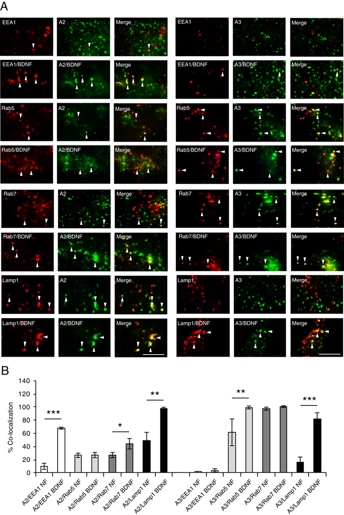
Figure 3.

EndophilinA2 and A3 are recruited to endosomes in response to BDNF. (A) Representative TIRF microscopy images of MEFs co-transfected with GFP-tagged EndophilinA2 (left panels) or EndophilinA3 (right panels) and the RFP-tagged endosomal markers EEA1, Rab5, Rab7 or Lamp1, in the presence and absence of 100 ng/ml BDNF. Arrows indicate colocalization of EndophilinA (green) with endosomal markers (red). (B) Quantitation of colocalization of transfected GFP-tagged EndophilinA2, and A3 with RFP-tagged EEA1, Rab5, Rab7 and Lamp1; n = 8–10 images per condition from 3 independent cell cultures, significance determined by unpaired two-tailed Student’s t-test test comparing non-stimulated to stimulated conditions, error = SEM, *p < 0.05, **p < 0.01, ***p < 0.001, scale bar = 10 µm.
