Figure 7.

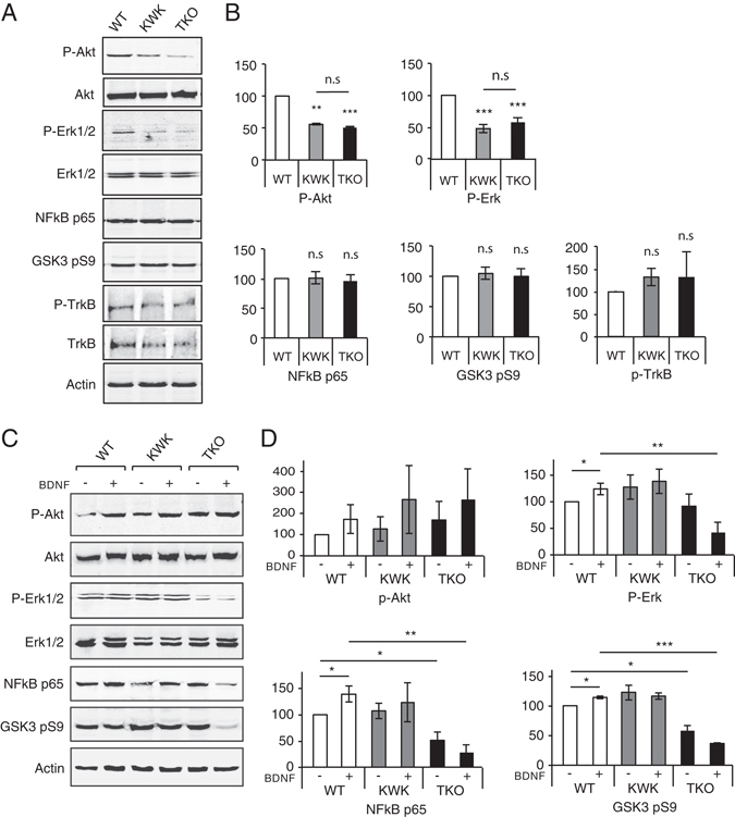
EndophilinA triple knockouts show changes in BDNF-mediated signaling. (A) Western blots of the indicated proteins in EndophilinA1/3 double knockout (KWK refers to Knockout, Wild-type, Knockout, for EndophilinA1, A2, A3), and EndophilinA triple knockout (TKO) brain lysates compared to wild-type. (B) Quantitation of P-Akt, P-Erk, NFkB p65, GSK3 pS9, and pTrkB protein levels in Western blots normalized to actin; n = 3 independent experiments, significance determined by unpaired two-tailed Student’s t-test comparing each condition to control, error = SEM, **p < 0.01, ***p < 0.001. (C) Western blots of the indicated proteins in EndophilinA1/3 double knockout (KWK refers to Knockout, Wild-type, Knockout, for EndophilinA1, A2, A3), and EndophilinA triple knockout (TKO) hippocampal cultures compared to wild-type in control and BDNF-treated conditions (100 ng/ml BDNF for 30 minutes). (D) Quantitation of P-Akt, P-Erk, NFkB p65 and GSK3 pS9 protein levels in Western blots normalized to actin; n = 3 independent experiments, significance determined by unpaired two-tailed Student’s t-test comparing each condition to control, error = SEM, **p < 0.01, ***p < 0.001.
