
Figure 6.
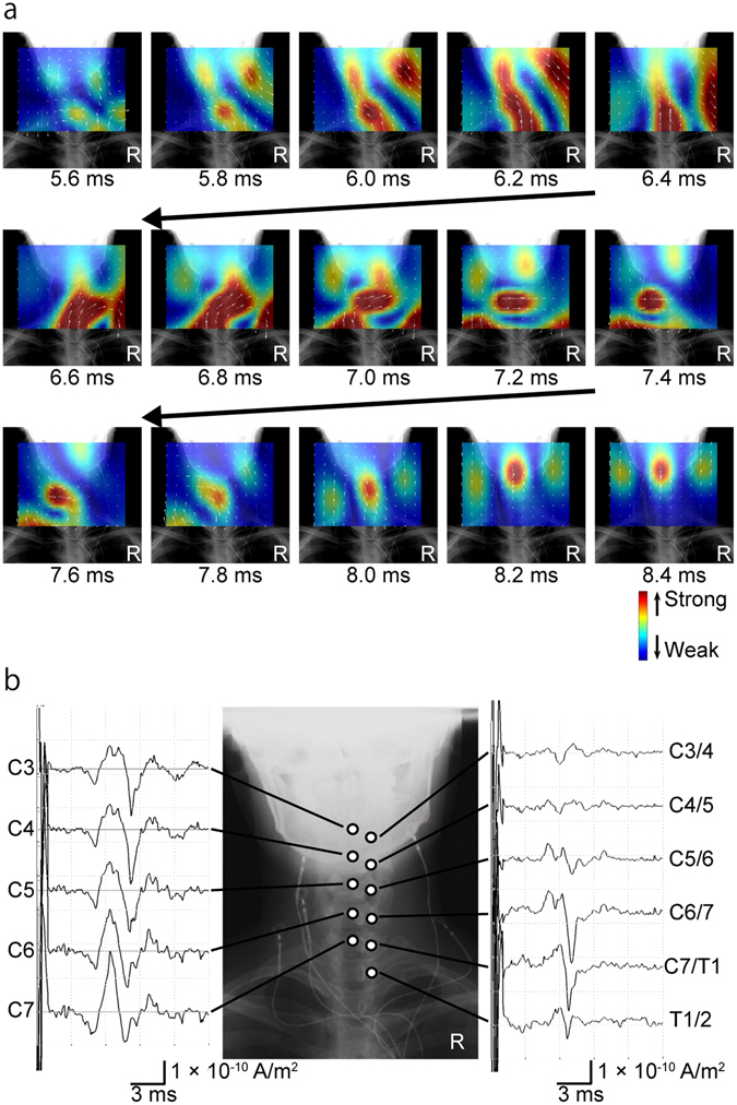
Reconstructed currents after stimulation of the right median nerve at the elbow. (a) Reconstructed current map. The currents flowed into the spinal canal from the right side and propagated cranially 5.6–6.4 ms post-stimulus. Rotated currents were observed 6.6–7.6 ms post-stimulus. Trailing currents propagated cranially 7.8–8.4 ms post-stimulus. (b) Reconstructed currents at the midline of the cervical spinal canal (C3 to C7) and at the adjacent intervertebral foramina (C3/4 to Th1/2). The first peak of the reconstructed currents at the midline of the cervical spinal canal did not propagate, but subsequent currents did (left graphs). Currents flowing into the intervertebral foramina were larger at C5/6 to C7/Th1 (right graphs).