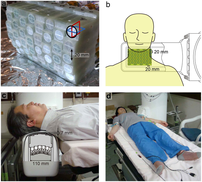
Figure 8.

The 120-channel superconducting quantum interference device (SQUID) biomagnetometer system used for the magnetospinography. (a) Individual SQUID sensors are arranged in an eight-by-five configuration at 20-mm intervals. Each sensor has three perpendicular pickup coils to detect magnetic fields in three orthogonal directions. (b) Overhead view of the sensor and subject as shown in Figs 1 and 5. (c) Cross-sectional image of the cryostat protrusion. The central grey area indicates the sensor array shown in (a). The sensors are arranged in the vertical direction and positioned in close contact with the upper inwall of the protrusion. The subject was in the supine position on a table with the posterior neck on the protrusion holding the SQUID sensor array inside. The upper surface of the protrusion is curved to fit the lordosis of the cervical spine. (d) Both median nerves were alternatively stimulated at the anterior of the elbow joint. Volar splints to suppress evoked movements are not shown here.
