
Figure 7.
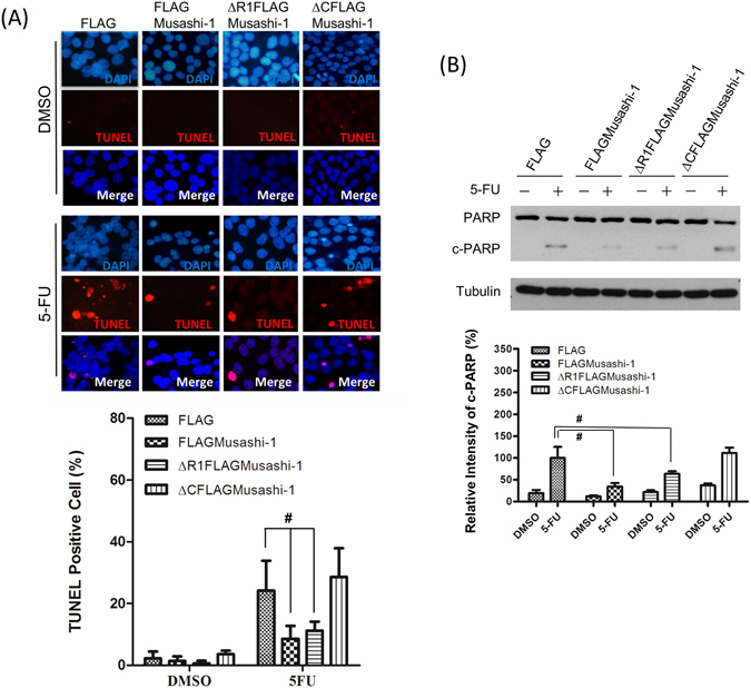
Musashi-1 inhibits 5-FU-induced apoptosis in HCT-116 cells. (A) Upper panel: FLAG, FLAGMusashi-1, ΔR1FLAGMusashi-1, and ΔCFLAGMusashi-1 HCT-116 stable clones were seeded on 22 mm × 22 mm coverslips and treated with 5-FU (400 μM) for 24 h. Cells were fixed in 4% paraformaldehyde and subjected to the TUNEL assay as described in the Methods. Lower panel: Statistical results of percentages of TUNEL positive cells. Error bars indicate the mean ± SD from three independent experiments. #p < 0.05. (B) Upper panel: FLAG, FLAGMusashi-1, ΔR1FLAGMusashi-1, and ΔCFLAGMusashi-1 HCT-116 stable clones were treated with or without 5-FU (400 μM) for 24 h. Total cellular proteins were isolated and subjected to immunoblotting with antibodies specific to PARP. Increased cleaved PARP (c-PARP) signals were observed in ΔCFLAGMusashi-1 HCT-116 stable clones. Tubulin indicates Tubulin protein as an internal control. Lower panel: Statistical results of percentages of relative intensity of c-PARP. Error bars indicate the mean ± SD from three independent experiments. #p < 0.05.