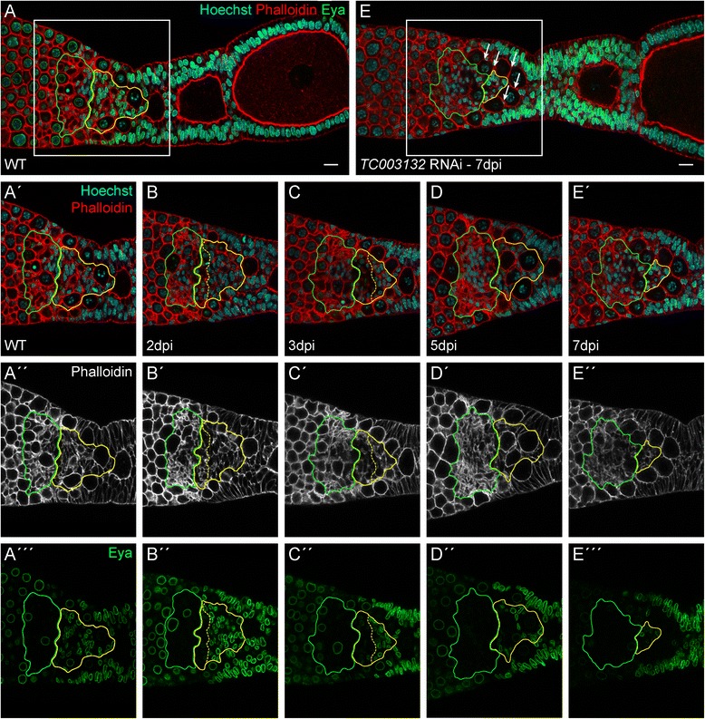
Fig. 4.

(A–A‴) Wild type ovariole stained with Phalloidin for f-Actin (red in A–A′, white in A″), Eya (green), and Hoechst (blue). Eya is expressed in central pre-follicular cells (CPC, yellow outline) and the somatic plug (SP, green outline) (B–E‴) Ovarioles dissected at indicated time points after adult knockdown of TC003132. A subset of CPC starts to express Eya at lower levels (border between high and low expression of Eya in CPCs is marked by the yellow dashed line) (B–B″). At 3dpi, CPCs occupy a smaller area and Eya expression is further reduced. The somatic plug (SP) appears less condensed and occupies a larger area (C–C″). CPCs expressing reduced amounts of Eya are lost at 5dpi (D–D″). The terminal phenotype can be observed at 7dpi. Only a small population of Eya positive CPCs is still present and the SP has expanded even more. Also encapsulation defects become apparent as young oocytes can be found in direct contact to each other (arrows, E–E‴). Scale bar: 10 μm
