
Fig. 3.
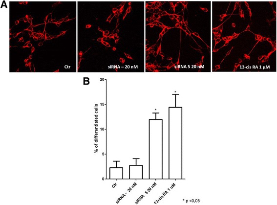
Analysis of siRNA DLL1 and 13-cis retinoic acid treated IMR-32 neuroblastoma cell line. a Confocal analysis of siRNA DLL1 and 13-cis retinoic acid in IMR-32 neuroblastoma cells after 3 days of transfection. β III tubulin antibody was used as neuron marker. b Morphometric analysis of cell differentiation induced by siRNA DLL1 and 13-cis retinoic acid. Neuronal differentiation was evaluated by measuring neurite length. The percentage of differentiated cells was calculated, considered as cells with neurites ≥50 μM in length in IMR-32 cells. *p < 0.05 vs control