Fig. 3.

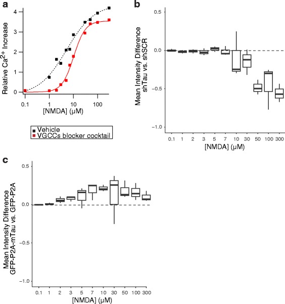
Tau modulates NMDAR-dependent Ca2+ influx. Live Ca2+ imaging was used to determine intracellular Ca2+ levels in WT neurons that were treated with different concentrations of NMDA at DIV14. a Representative relationship between NMDA doses and Ca2+ influx in neurons treated with vehicle or with a cocktail of voltage-gated calcium channel (VGCC) blockers for 30 min prior to and throughout NMDA application (see Methods). The VGCC blocker cocktail isolates NMDAR-dependent Ca2+ influx. Increases in fluorescence signals are expressed relative to baseline measurements obtained in the same wells. b, c In neurons treated with the VGCC blocker cocktail, tau knockdown decreased b, whereas tau overexpression increased c NMDAR-dependent Ca2+ influx. The boxplots represent the distribution of the differences in mean fluorescence between shTau- vs. shSCR-expressing neurons in b and GFP-P2A-mTauWT vs. GFP-P2A expressing neurons in c at each dose across independent experiments. Numbers of independent experiments (n) with cumulative well numbers per condition in parentheses: a 1 (3), b 3 (9), and c 3 (9). When comparing mean differences across all doses within any given panel, a one-sided, one-sample t-test revealed significant differences between experimental and control conditions in (b: P < 0.05) and (c: P < 0.05)
